Dr Grand Roman Joldes
Towards translating the benefits of patient specific biomechanics into clinical practice
Dr Grand Roman Joldes from the Faculty of Engineering and Mathematical Sciences, The University of Western Australia, was awarded a 2016 Raine Priming Grant.
Dr Joldes is a Senior Research Fellow in the Department of Mechanical Engineering within the Faculty of Engineering and Mathematical Sciences at The University of Western Australia. He completed his PhD with Distinction in 2010 and was awarded a Raine Priming Grant to develop algorithms that allow generation of computational models for medical images, as well as robust, fast and accurate solution methods.
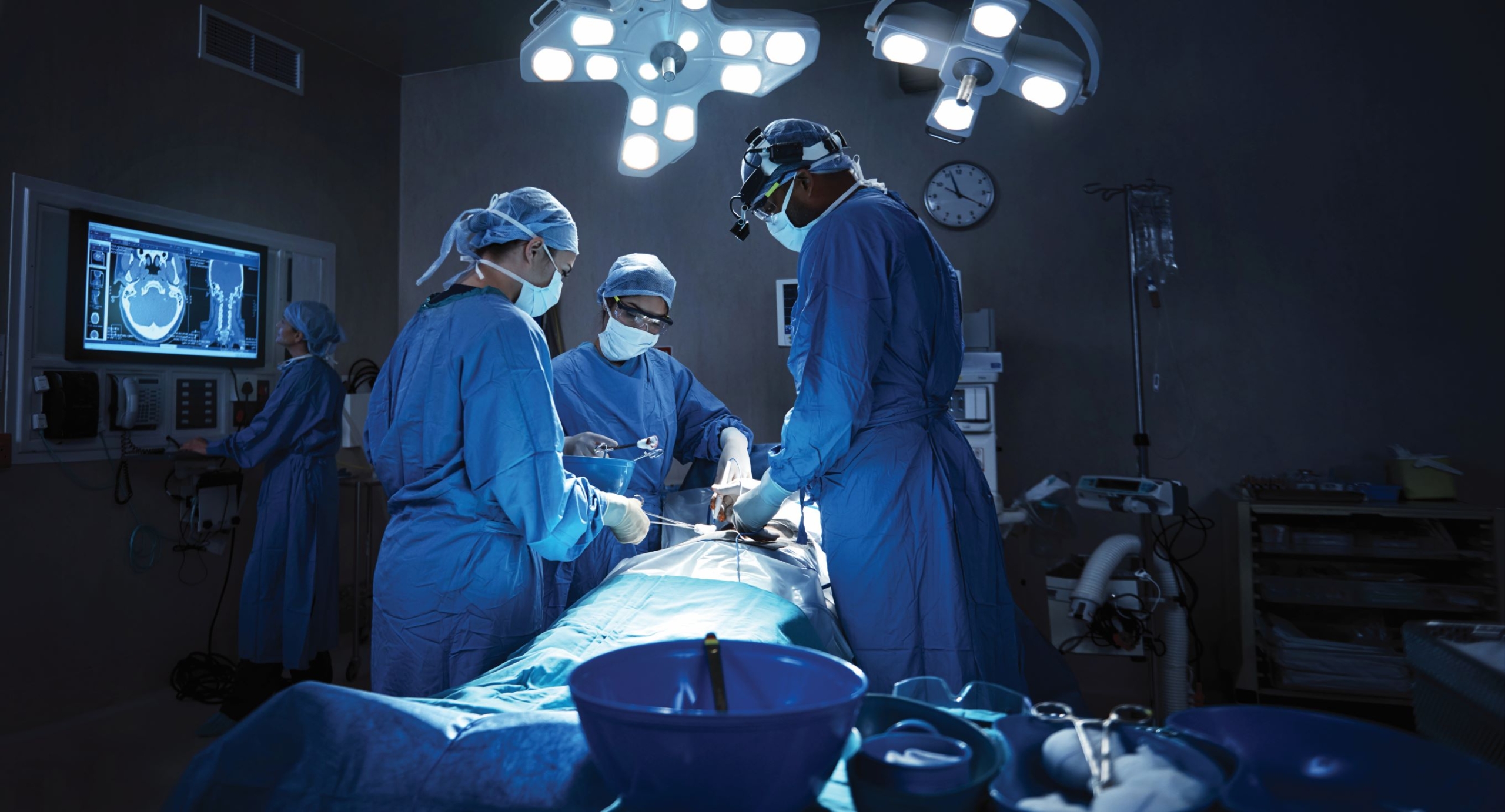
There is widespread international concern about the cost of meeting rising expectations for healthcare, particularly if large numbers of people require currently expensive procedures such as brain surgery. The costs can be reduced by using improved machinery to help surgeons perform these procedures quickly and accurately with minimal adverse effects. A novel partnership between surgeons and machines, made possible by advances in computing and engineering technology, could overcome many of the limitations of traditional surgery. The development of computational algorithms for patient-specific biomechanical modelling has continued during the last 12 months, where these algorithms have been used to solve clinically relevant problems such as brain deformation during electrode implantation for epilepsy treatment and blood flow within abdominal aortic aneurysms.
This study has led to four publications in 2018, supported two successful NHMRC grant applications, and Dr Joldes receiving a UWA Research Collaboration Award that will facilitate continued collaborations with Harvard Medical School. The findings from this project have the potential to transform current methods and result in great improvements into tools that deliver real outcomes for patients.
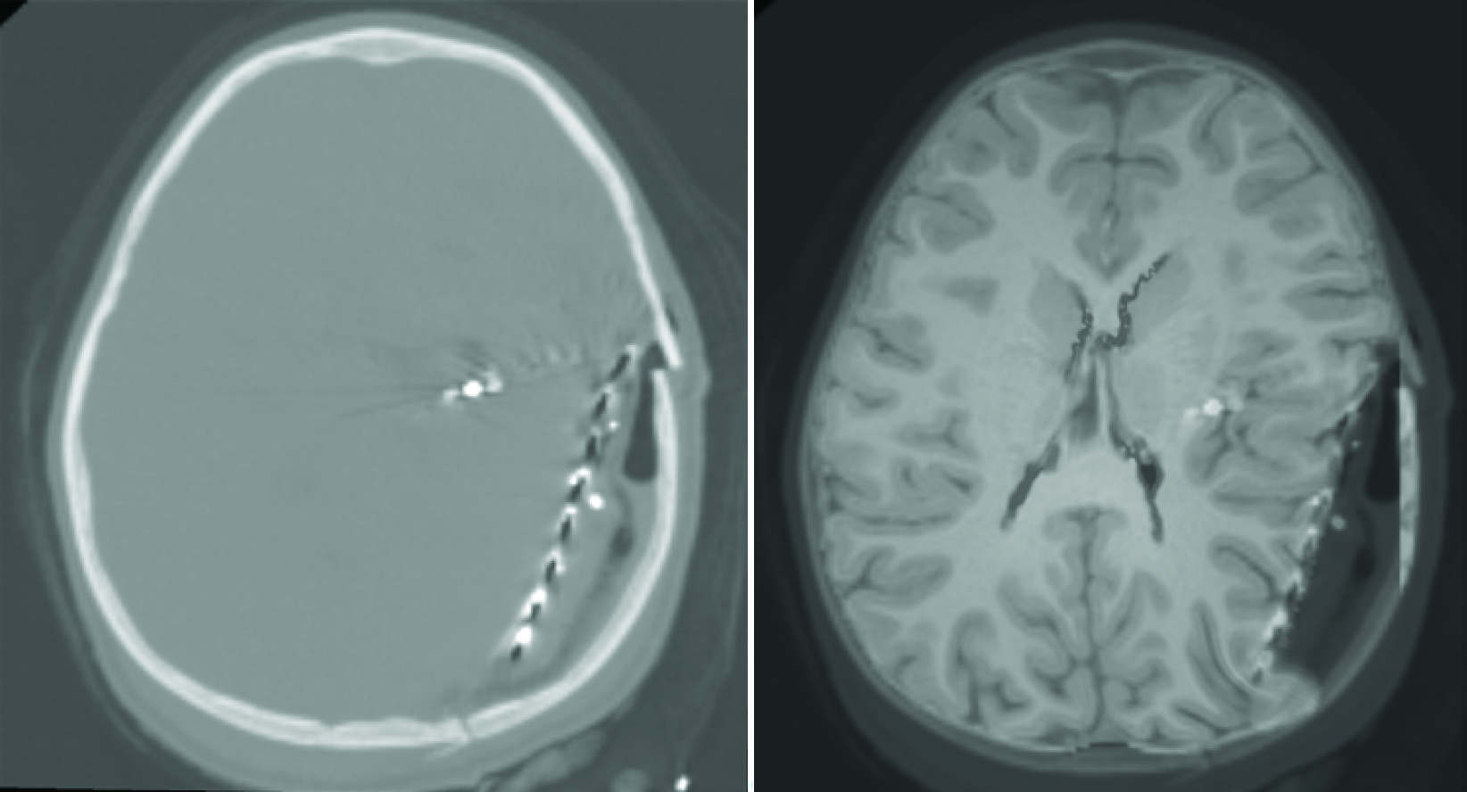
Left panel: CT brain image after implantation of electrodes for epilepsy treatment. No details regarding brain structures can be distinguished from this image. MRI imaging is not possible due to the presence of metal electrodes.
Right panel: Preoperative MRI image deformed using a biomechanical model using the displacement introduced by electrodes as input. Computations performed using the algorithms developed by Dr Joldes. The medical data was provided by Harvard Medical School.